Anatomy and biomechanics of the foot
It is often considered that the basic function of the human foot is to provide firm foundation for upright standing. From evolutionary point of view, the foot has been developing as a dynamic mechanism and is therefore not quite adapt for standing in one position, as it causes fatigue and pain. We prefer, and are biologically predetermined to be walking as opposed to sitting or standing.
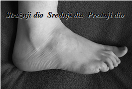
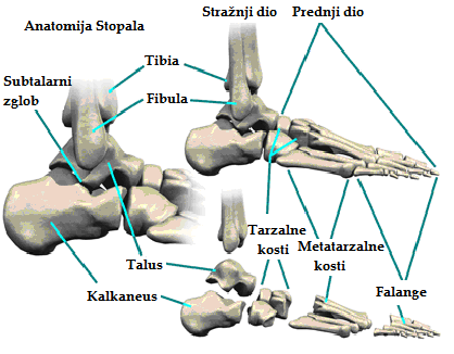
As a unique functional system, the human foot behaves like an elastic feather, which is of great importance for support and propulsion in the walking cycle. Anatomically, the foot can be divided into three parts. Hindfoot is composed of bones called Talus and Calcaneus, Middlefoot contains Navicular, Cuboid and three Cuneiforms, while Forefoot is made of five Metatarsal bones, fourteen Phalanges and two Sesamoid bones which are all strongly bonded by muscles, tendons, ligaments, peripheral circulation streams and nerves. This structure facilitates collective and interdependent movement of all anatomical elements in the foot.
During a walk, bodyweight is first being partly transmitted from shin to Talus (ankle bone), and then mostly to Calcaneus and Forefoot area, creating three points of pressure. Those three points are Tuber of Calcaneus (1), the head of V Metatarsal bone (2), and the head of I Metatarsal bone (3). These points, connected by bones, ligaments and muscles form two longitudinal (inner and outer) and two transversal (front and back) arches of the foot.
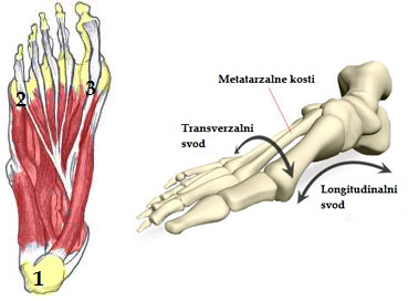
Longitudinal arches start from Calcaneus and stretch forward towards Phalanges. Inner arch starts at Calcaneus and stretches over Talus, Navicular, first Cuneiform and across I Metatarsal bone to end on it's head. The outer longitudinal arch stretches over Cuboid and V Metatarsal bone.
The highest point of the inner arch is the Navicular bone (15 mm from the ground), and highest outer arch point is the Cuboid bone (5 mm from the ground). Front transversal arch connects the heads of I-V Metatarsal to the highest point in the head of II Metatarsal. Back transversal arch is located over three Cuneiforms and a Cuboid bone.
Statics and dynamics of the foot and arch support are all conditioned by the proper muscular function, and the functions of ligaments which connect the bones. Most important ligaments in this respect are lig. calcaneonaviculare - LC, lig. plantare longum - LP and aponeurosis plantaris - AP.
The arches of the foot form while the child is still in mother's womb, but they are clearly visible only after reaching the age of about two years, when the fat pads on the child's soles get thinner.
Walking in a an upright position is one of the main characteristics of the human species, and although we all basically walk in the same manner, there are minor individual differences that allow us to recognize friends and acquaintances even from relatively great distances.